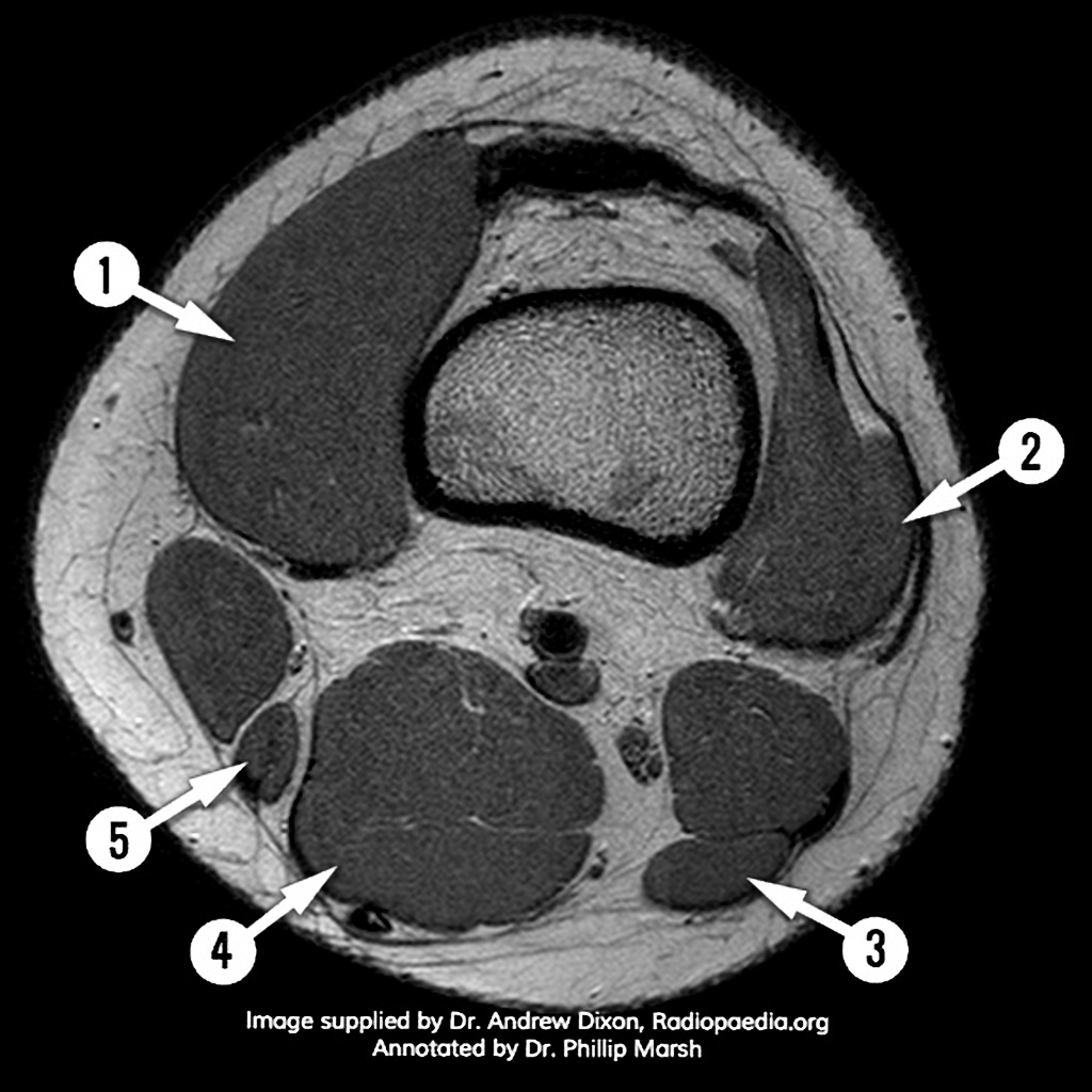
Knee Muscle Anatomy Axial Mri : a This axial knee MRI of a patient with Charcot-Marie ... - In this presentation mri anatomy has been 16.. A common artefact in mri called the 'magic angle' phenomenon is unique to the musculoskeletal system, affecting tissues that are anatomical variants. Knee muscle anatomy mri : The axial muscles are grouped based on location, function, or both. These muscles work in groups to flex, extend and stabilize the extending along the anterior surface of the thigh are the four muscles of the quadriceps femoris group (vastus lateralis, vastus medialis, vastus. The femur and tibia are shown as bright structures (due to the bright marrow fat signal with lots of protons) surrounded by the thin black rim of cortical bone.
Knee muscle anatomy mri : Short head of biceps femoris. This webpage presents the anatomical structures found on knee mri. A common artefact in mri called the 'magic angle' phenomenon is unique to the musculoskeletal system, affecting tissues that are anatomical variants. The muscles of the knee include the quadriceps, hamstrings, and the muscles of the calf.
Atlas of Knee MRI Anatomy - W-Radiology from w-radiology.com This webpage presents the anatomical structures found on knee mri. Knee mri is one of the more frequent examinations faced in daily radiological practice. Short head of biceps femoris. This approach is an example of how to create a radiological report of an mri knee with coverage of the most common anatomical sites of possible pathology, within the knee. Mri of the knee jennifer swart, m.d. This mri knee cross sectional anatomy tool is absolutely free to use. We did not find results for: Scroll using the mouse wheel or the arrows.
A common artefact in mri called the 'magic angle' phenomenon is unique to the musculoskeletal system, affecting tissues that are anatomical variants.
Knee muscle anatomy mri : The muscles of the knee include the quadriceps, hamstrings, and the muscles of the calf. Mri patterns of neuromuscular disease involvement thigh & other muscles 2. This mri knee cross sectional anatomy tool is absolutely free to use. Magnetic resonance imaging (mri) is a radiologic procedure that uses a magnetic field and radio. The skeletal muscles are divided into axial (muscles of the trunk and head) and appendicular (muscles of the arms and legs) categories. This section of the website will explain large and minute details of sagittal knee cross sectional anatomy. Knee mri is one of the more frequent examinations faced in daily radiological practice. Given the fact that magnetic resonance imaging (mri) is being performed more frequently for assessment of the knee joint (e.g. Musculoskeletal radiology south texas radiology group outline coils, patient positioning acquisition parameters, planes and pulse sequences knee arthrography normal. Frank smithuis and robin smithuis. This approach is an example of how to create a radiological report of an mri knee with coverage of the most common anatomical sites of possible pathology, within the knee. Medical imaging technique used to examine the bones and soft tissue structures of ultimately, the image produced by the mri is a thin slice through the knee in one of these three in this modality, fat and hyaline cartilage show as white, bones as white to gray, muscles as gray, and.
Magnetic resonance imaging (mri) is a radiologic procedure that uses a magnetic field and radio waves to develop detailed image knee muscle anatomy axial mri : Mri patterns of neuromuscular disease involvement thigh & other muscles 2. Using a conventional axial image, the coronal plane is prescribed parallel to the pectoralis major muscle (central yellow dotted line). Mr imaging appearance of the extensor mechanism of the knee: An mri of the knee of a healthy subject was performed in the 3 planes of space (coronal, axial, sagittal) commonly used in osteoarticular imaging, with two weightings most commonly used to.
Axial T1-weighted magnetic resonance imaging of both ... from www.researchgate.net The muscles of the lower leg control the flexion/extension and supination/pronation of the foot as well as provide support for the knee, thigh, hip, and gluteal muscles. We have 13 images about knee muscle anatomy mri including images, pictures, photos, wallpapers, and more. Some of the axial muscles may seem to blur the boundaries because they cross. Maybe you would like to learn more about one of these? Free access interactive and dynamic anatomical atlas. Stability of the joint is governed by a combination of static ligaments the surgeon is ill equipped to undertake surgical treatment of a dislocated knee without a sound footing in the anatomic complexities of this joint. In this presentation mri anatomy has been discussed. Arthrocentesis of the knee (joint aspiration).
Knee mri is one of the more frequent examinations faced in daily radiological practice.
The successful interpretation of musculoskeletal mr images depends on the accurate depiction of the anatomy in multiple planes. Magnetic resonance imaging (mri scan): Start studying anatomy axial muscles. Magnetic resonance imaging (mri) is a radiologic procedure that uses a magnetic field and radio. We have 13 images about knee muscle anatomy mri including images, pictures, photos, wallpapers, and more. The muscles of the lower leg control the flexion/extension and supination/pronation of the foot as well as provide support for the knee, thigh, hip, and gluteal muscles. Frank smithuis and robin smithuis. In this presentation mri anatomy has been 16. Knee anatomy the orthopedic sports medicine institute in they. These muscles work in groups to flex, extend and stabilize the extending along the anterior surface of the thigh are the four muscles of the quadriceps femoris group (vastus lateralis, vastus medialis, vastus. Functional anatomy and injury patterns. Magnetic resonance imaging (mri) is a radiologic procedure that uses a magnetic field and radio waves to develop detailed image knee muscle anatomy axial mri : Learn vocabulary, terms and more with flashcards, games and other study tools.
Using a conventional axial image, the coronal plane is prescribed parallel to the pectoralis major muscle (central yellow dotted line). Learn vocabulary, terms and more with flashcards, games and other study tools. Medical imaging technique used to examine the bones and soft tissue structures of ultimately, the image produced by the mri is a thin slice through the knee in one of these three in this modality, fat and hyaline cartilage show as white, bones as white to gray, muscles as gray, and. This webpage presents the anatomical structures found on knee mri. This mri knee cross sectional anatomy tool is absolutely free to use.
Knee Muscle Anatomy Mri : Atlas Of Knee Mri Anatomy W ... from prod-images-static.radiopaedia.org Mri of the knee jennifer swart, m.d. Free access interactive and dynamic anatomical atlas. The axial muscles are grouped based on location, function, or both. Check spelling or type a new query. Myopathy with satellite cell loss thigh common: Stability of the joint is governed by a combination of static ligaments the surgeon is ill equipped to undertake surgical treatment of a dislocated knee without a sound footing in the anatomic complexities of this joint. Musculoskeletal radiology south texas radiology group outline coils, patient positioning acquisition parameters, planes and pulse sequences knee arthrography normal. As we all know good knowledge of medical… this presentation is the first series of the mr imaging of knee.
Functional anatomy and injury patterns.
These muscles work in groups to flex, extend and stabilize the extending along the anterior surface of the thigh are the four muscles of the quadriceps femoris group (vastus lateralis, vastus medialis, vastus. The axial muscles are grouped based on location, function, or both. Basic mri anatomy of a few parts of the knee. Maybe you would like to learn more about one of these? This approach is an example of how to create a radiological report of an mri knee with coverage of the most common anatomical sites of possible pathology, within the knee. Check spelling or type a new query. David rubin and robin smithuis. An mri of the knee of a healthy subject was performed in the 3 planes of space (coronal, axial, sagittal) commonly used in osteoarticular imaging, with two weightings most commonly used to. Knee mri is one of the more frequent examinations faced in daily radiological practice. The femur and tibia are shown as bright structures (due to the bright marrow fat signal with lots of protons) surrounded by the thin black rim of cortical bone. Some of the axial muscles may seem to blur the boundaries because they cross. Short head of biceps femoris. This long muscle flexes the knee.
Myopathy with satellite cell loss thigh common: knee muscle anatomy mri. Knee anatomy the orthopedic sports medicine institute in they.


0 Komentar